Image:SEM blood cells.jpg
From Wikipedia, the free encyclopedia
Size of this preview: 385 × 479 pixels
Full resolution (1,800 × 2,239 pixels, file size: 1.33 MB, MIME type: image/jpeg)
| |
This is a file from the Wikimedia Commons. The description on its description page there is shown below. |
Summary
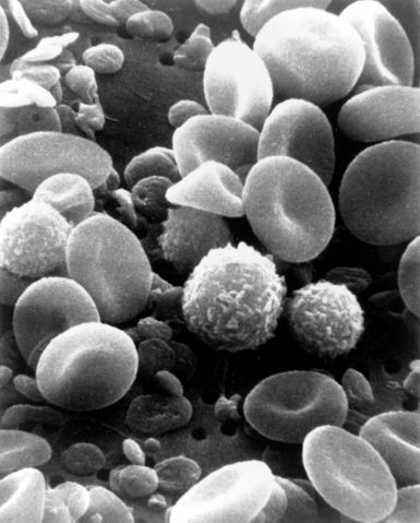
| Description |
This is a scanning electron microscope image from normal circulating human blood. One can see red blood cells, several white blood cells including lymphocytes, a monocyte, a neutrophil, and many small disc-shaped platelets. Red cells are nonnucleated and contain hemoglobin, an important protein which contains iron and allows the cell to carry oxygen to other parts of the body. They also carry carbon dioxide away from the lungs. The infection-fighting white blood cells are classified in two main groups: granular and agranular. Granulocytes are formed in bone marrow; agranulocytes are produced by lymph nodes and spleen. There are two types of agranulocytes: lymphocytes, which fight disease by producing antibodies and thus destroying foreign material, and monocytes. Platelets are tiny cells formed in bone marrow and are necessary for blood clotting. Type: Black & White Print |
|---|---|
| Source |
Image and description: National Cancer Institute |
| Date |
Date Created: February 1982 |
| Author |
Bruce Wetzel (photographer). Harry Schaefer (photographer) |
| Permission ( Reusing this image) |
"All images are in the public domain and may be used, linked, or reproduced without permission. If an image is used, credit should be given to the listed source and/or author."
|
Licensing
 |
This work is in the public domain in the United States because it is a work of the United States Federal Government under the terms of Title 17, Chapter 1, Section 105 of the US Code. See Copyright. Note: This only applies to works of the Federal Government and not to the work of any individual U.S. state, territory, commonwealth, county, municipality, or any other subdivision. This template also does not apply to postage stamps published by the United States Postal Service after 1978. (See 206.02(b) of Compendium II: Copyright Office Practices). العربية | Български | Česky | Deutsch | English | Español | Français | Magyar | Italiano | 日本語 | 한국어 | Polski | Português | 中文(繁體) | 中文(简体) | +/- |
 |
File history
Click on a date/time to view the file as it appeared at that time.
| Date/Time | Dimensions | User | Comment | |
|---|---|---|---|---|
| current | 20:27, 7 October 2006 | 1,800×2,239 (1.33 MB) | DO11.10 | |
| 03:00, 4 October 2006 | 1,800×2,239 (989 KB) | DO11.10 | ({{Information |Description=This is a scanning electron microscope image from normal circulating human blood. One can see red blood cells, several white blood cells including lymphocytes, a monocyte, a neutrophil, and many small disc-shaped platelets. Red ) | |
| 01:09, 4 October 2006 | 500×326 (36 KB) | DO11.10 | ({{Information |Description= A three-dimensional ultrastructural image analysis of a T-lymphocyte (right), a platelet (centre) and a red blood cell (left), using a Hitachi S-570 scanning electron microscope (SEM) equipped with a GW Backscatter Detector. ) |