Image:FMRI.jpg
From Wikipedia, the free encyclopedia
No higher resolution available.
FMRI.jpg (250 × 208 pixels, file size: 11 KB, MIME type: image/jpeg)
| |
This is a file from the Wikimedia Commons. The description on its description page there is shown below. |
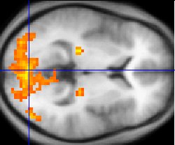
Sample fMRI data
Source: http://en.wikipedia.org/wiki/Image:FMRI.jpg
Public-domain: copyright disclaimed Washington irving] 07:49, 8 Mar 2004 (UTC)
This example of fMRI data shows regions of activation including primary visual cortex (V1, BA17), extrastriate visual cortex and lateral geniculate body in a comparison between a task involving a complex moving visual stimulus and rest condition (viewing a black screen). The activations (yellow-red) are shown (as is typical) against a background based on the average structural images from the subjects in the experiment.
 |
This image has been (or is hereby) released into the public domain by its author, Washington irving at the English Wikipedia project. This applies worldwide. In case this is not legally possible: |
File history
Click on a date/time to view the file as it appeared at that time.
| Date/Time | Dimensions | User | Comment | |
|---|---|---|---|---|
| current | 00:52, 9 December 2004 | 250×208 (11 KB) | Superborsuk | (Sample fMRI data This example of fMRI data shows regions of activation including primary visual cortex (V1, BA17), extrastriate visual cortex and lateral geniculate body in a comparison between a task involving a complex moving visual stimulus and re) |