Retina
2008/9 Schools Wikipedia Selection. Related subjects: Health and medicine
| Retina | |
|---|---|
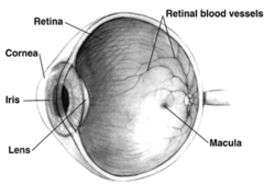
| Right human eye cross-sectional view. Courtesy NIH National Eye Institute. Many animals have eyes different from the human eye. | |
| Gray's | subject #225 1014 |
| Artery | central retinal artery |
| MeSH | Retina |
| Dorlands/Elsevier | r_10/12705919 |
The vertebrate retina is the light sensitive inner layer of the eye. Two of its three types of photoreceptor cells, rods and cones, receive light and transform it into image-forming signals which are transmited through the optic nerve to the brain. In this respect, the retina is comparable to the film in a camera.
The third and more recently discovered category of photosensitive cells is probably not involved in image-forming vision. These are a small proportion, about 2% in humans, of the retina's ganglion cells, themselves photosensitive through the photopigment melanopsin, which transmit information about light through the RHT (retinohypothalamic tract) directly to the SCN (suprachiasmatic nucleus) and other brain structures. Signals from these ganglion cells are used to adjust the size of the pupil, entrain the body's circadian rhythms and acutely suppress the pineal hormone melatonin, processes which in fact function in many blind people who do not have functioning rods and cones.
While rods and cones respond maximally to wavelengths around 555 nanometers (green), the light sensitive ganglion cells respond maximally to about 480nm (blue-violet). There are several different photopigments involved.
Neural signals from the rods and cones undergo complex processing by other neurons of the retina. The output takes the form of action potentials in retinal ganglion cells whose axons form the optic nerve. Several important features of visual perception can be traced to the retinal encoding and processing of light.
In vertebrate embryonic development, the retina and the optic nerve originate as outgrowths of the developing brain. Hence, the retina is part of the central nervous system (CNS). It is the only part of the CNS that can be imaged directly.
The unique structure of the blood vessels in the retina has been used for biometric identification.
Anatomy of vertebrate retina
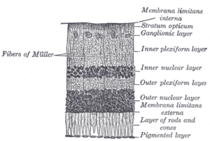
The vertebrate retina has ten distinct layers. From innermost to outermost, they include:
- Inner limiting membrane - Müller cell footplates
- Nerve fibre layer
- Ganglion cell layer - Layer that contains nuclei of ganglion cells and gives rise to optic nerve fibers.
- Inner plexiform layer
- Inner nuclear layer
- Outer plexiform layer - In the macular region, this is known as the Fibre layer of Henle.
- Outer nuclear layer
- External limiting membrane - Layer that separates the inner segment portions of the photoreceptors from their cell nuclei.
- Photoreceptor layer - Rods / Cones
- Retinal pigment epithelium
Physical structure of human retina
In adult humans the entire retina is 72% of a sphere about 22 mm in diameter. An area of the retina is the optic disc, sometimes known as "the blind spot" because it lacks photoreceptors. It appears as an oval white area of 3 mm². Temporal (in the direction of the temples) to this disc is the macula. At its centre is the fovea, a pit that is most sensitive to light and is responsible for our sharp central vision. Human and non-human primates possess one fovea as opposed to certain bird species such as hawks who actually are bifoviate and dogs and cats who possess no fovea but a central band known as the visual streak. Around the fovea extends the central retina for about 6 mm and then the peripheral retina. The edge of the retina is defined by the ora serrata. The length from one ora to the other (or macula), the most sensitive area along the horizontal meridian is about 3.2 mm.
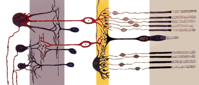
In section the retina is no more than 0.5 mm thick. It has three layers of nerve cells and two of synapses. The optic nerve carries the ganglion cell axons to the brain and the blood vessels that open into the retina. As a byproduct of evolution, the ganglion cells lie innermost in the retina while the photoreceptive cells lie outermost. Because of this arrangement, light must first pass through the thickness of the retina before reaching the rods and cones. However it does not pass through the epithelium or the choroid (both of which are opaque).
The white blood cells in the capillaries in front of the photoreceptors can be perceived as tiny bright moving dots when looking into blue light. This is known as the blue field entoptic phenomenon (or Scheerer's phenomenon).
Between the ganglion cell layer and the rods and cones there are two layers of neuropils where synaptic contacts are made. The neuropil layers are the outer plexiform layer and the inner plexiform layer. In the outer the rod and cones connect to the vertically running bipolar cells and the horizontally oriented horizontal cells connect to ganglion cells.
The central retina is cone-dominated and the peripheral retina is rod-dominated. In total there are about seven million cones and a hundred million rods. At the centre of the macula is the foveal pit where the cones are smallest and in a hexagonal mosaic, the most efficient and highest density. Below the pit the other retina layers are displaced, before building up along the foveal slope until the rim of the fovea or parafovea which is the thickest portion of the retina. The macula has a yellow pigmentation from screening pigments and is known as the macula lutea.
Vertebrate and cephalopod retina differences
The vertebrate retina is inverted in the sense that the light sensing cells sit at the back side of the retina, so that light has to pass through a layer of neurons before it reaches the rods and cones. By contrast, the cephalopod retina is everted: the photoreceptors are located at the front side of the retina, with processing neurons behind them. Because of this, cephalopods do not have a blind spot.
The cephalopod retina does not originate as an outgrowth of the brain, as the vertebrate one does. This shows that vertebrate and cephalopod eyes are not homologous but have evolved separately.
Physiology
An image is produced by the "patterned excitation" of the cones and rods in the retina. The excitation is processed by the neuronal system and various parts of the brain working in parallel to form a representation of the external environment in the brain.
The cones respond to bright light and mediate high-resolution vision and colour vision. The rods respond to dim light and mediate lower-resolution, black-and-white, night vision. It is a lack of cones sensitive to red, blue, or green light that causes individuals to have deficiencies in colour vision or various kinds of colour blindness. Humans and old world monkeys have three different types of cones ( trichromatic vision) while other mammals lack cones with red sensitive pigment and therefore have poorer (dichromatic) colour vision.
When light falls on a receptor it sends a proportional response synaptically to bipolar cells which in turn signal the retinal ganglion cells. The receptors are also 'cross-linked' by horizontal cells and amacrine cells, which modify the synaptic signal before the ganglion cells. Rod and cone signals are intermixed and combine, although rods are mostly active in very poorly lit conditions and saturate in broad daylight, while cones function in brighter lighting because they are not sensitive enough to work at very low light levels.
Despite the fact that all are nerve cells, only the retinal ganglion cells and few amacrine cells create action potentials. In the photoreceptors, exposure to light hyperpolarizes the membrane in a series of graded shifts. The outer cell segment contains a photopigment. Inside the cell the normal levels of cGMP keeps the Na+ channel open and thus in the resting state the cell is depolarised. The photon causes the retinal bound to the receptor protein to isomerise to trans-retinal. This causes receptor to activate multiple G-proteins. This in turn causes the Ga-subunit of the protein to bind and degrade cGMP inside the cell which then cannot bind to the CNG Na+ channels. Thus the cell is hyperpolarised. The amount of neurotransmitter released is reduced in bright light and increases as light levels fall. The actual photopigment is bleached away in bright light and only replaced as a chemical process, so in a transition from bright light to darkness the eye can take up to thirty minutes to reach full sensitivity (see dark adaptation).
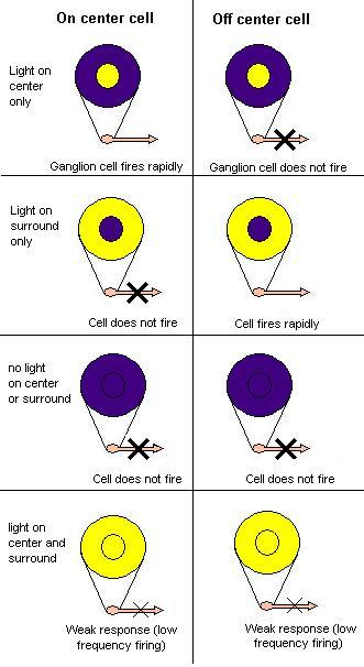
In the retinal ganglion cells there are two types of response, depending on the receptive field of the cell. The receptive fields of retinal ganglion cells comprise a central approximately circular area, where light has one effect on the firing of the cell, and an annular surround, where light has the opposite effect on the firing of the cell. In ON cells, an increment in light intensity in the centre of the receptive field causes the firing rate to increase. In OFF cells, it makes it decrease. In a linear model, this response profile is well described by a Difference of Gaussians and is the basis for edge detection algorithms. Beyond this simple difference ganglion cells are also differentiated by chromatic sensitivity and the type of spatial summation. Cells showing linear spatial summation are termed X cells (also called "parvocellular", "P", or "midget" ganglion cells), and those showing non-linear summation are Y cells (also called "magnocellular, "M", or "parasol" retinal ganglion cells), although the correspondence between X and Y cells (in the cat retina) and P and M cells (in the primate retina) is not as simple as it once seemed.
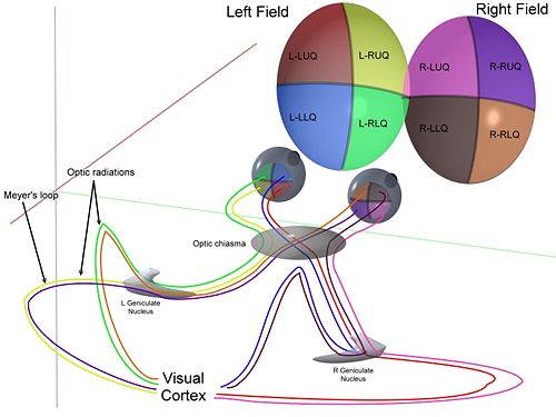
In the transfer of visual signals to the brain, the visual pathway, the retina is vertically divided in two, a temporal (nearer to the temple) half and a nasal (nearer to the nose) half. The axons from the nasal half cross the brain at the optic chiasma to join with axons from the temporal half of the other eye before passing into the lateral geniculate body.
Although there are more than 130 million retinal receptors, there are only approximately 1.2 million fibres (axons) in the optic nerve; a large amount of pre-processing is performed within the retina. The fovea produces the most accurate information. Despite occupying about 0.01% of the visual field (less than 2° of visual angle), about 10% of axons in the optic nerve are devoted to the fovea. The resolution limit of the fovea has been determined at around 10,000 points. The information capacity is estimated at 500,000 bits per second (for more information on bits, see information theory) without colour or around 600,000 bits per second including colour.
Spatial Encoding
The retina, unlike a camera, does not simply send a picture to the brain. The retina spatially encodes (compresses) the image to fit the limited capacity of the optic nerve. Compression is necessary because there are 100 times more Photoreceptor cells than ganglion cells as mentioned above. The retina does so by "decorrelating" the incoming images in a manner to be described below. These operations are carried out by the centre surround structures as implemented by the bipolar and ganglion cells.
There are two types of center surround structures in the retina -- on-centers and off-centers. On-centers have a positively weighted centre and a negatively weighted surround. Off-centers are just the opposite. Positive weighting is more commonly known as excitatory and negative weighting is more commonly known as inhibitory.
These center surround structures are not physical in the sense that you cannot see them by staining samples of tissue and examining the retina's anatomy. The centre surround structures are logical (i.e., mathematically abstract) in the sense that they depend on the connection strengths between ganglion and bipolar cells. It is believed that the connection strengths between cells is caused by the number and types of ion channels embedded in the synapses between the ganglion and bipolar cells. Stephen Kuffler in the 1950s was the first person to begin to understand these centre surround structures in the retina of cats. See Receptive field for figures and more information on centre surround structures. See chapter 3 of David Hubel's on-line book (listed below) for an excellent introduction.
The centre surround structures are mathematically equivalent to the edge detection algorithms used by computer programmers to extract or enhance the edges in a digital photograph. Thus the retina performs operations on the image to enhance the edges of objects within its visual field. For example, in a picture of a dog, a cat and a car, it is the edges of these objects that contain the most information. In order for higher functions in the brain (or in a computer for that matter) to extract and classify objects such as a dog and a cat, the retina is the first step to separating out the various objects within the scene.
As an example, the following matrix is at the heart of the computer algorithm that implements edge detection. This matrix is the computer equivalent to the center surround structure. In this example, each box (element) within this matrix would be connected to one photoreceptor. The photoreceptor in the center is the current receptor being processed. The center photoreceptor is multiplied by the +1 weight factor. The surrounding photoreceptors are the "nearest neighbors" to the centre and are multiplied by the -1/8 value. The sum of all nine of these elements is finally calculated. This summation is repeated for every photoreceptor in the image by shifting left to the end of a row and then down to the next line.
The total sum of this matrix is zero if all the inputs from the nine photoreceptors are the same value. The zero result indicates the image was uniform (non-changing) within this small patch. Negative or positive sums mean something was varying (changing) within this small patch of nine photoreceptors.
| -1/8 | -1/8 | -1/8 |
| -1/8 | +1 | -1/8 |
| -1/8 | -1/8 | -1/8 |
The above matrix is only an approximation to what really happens inside the retina. First, the table is square while the centre surround structures in the retina are circular. Second, neurons operate on spike trains traveling down nerve cell axons. Computers operate on a single constant number from each input pixel (the computer equivalent of a photoreceptor). Third, the retina performs all these calculations in parallel while the computer operates on each pixel one at a time. There are no repeated summations and shifting as there would be in a computer. Forth, the horizontal and amacrine cells play a significant role in this process but that is not represented here.
Here is an example of an input image and how edge detection would modify it.
Once the image is spatially encoded by the centre surround structures, the signal is sent out the optical nerve (via the axons of the ganglion cells) through the optic chiasm to the LGN ( lateral geniculate nucleus). The exact function of the LGN is unknown at this time. The output of the LGN is then sent to the back of the brain. Specifically the output of the LGN "radiates" out to the V1 Primary visual cortex.
Simplified Signal Flow: Photoreceptors ==> Bipolor ==> Ganglion ==> Chiasm ==> LGN ==> V1 cortex
Diseases and disorders
There are many inherited and acquired diseases or disorders that may affect the retina. Some of them include:
- Retinitis pigmentosa is a group of genetic diseases that affect the retina and causes the loss of night vision and peripheral vision.
- Macular degeneration describes a group of diseases characterized by loss of central vision because of death or impairment of the cells in the macula.
- Cone-rod dystrophy (CORD) describes a number of diseases where vision loss is caused by deterioration of the cones and/or rods in the retina.
- In retinal separation, the retina detaches from the back of the eyeball. Ignipuncture is an outdated treatment method.
- Both hypertension and diabetes mellitus can cause damage to the tiny blood vessels that supply the retina, leading to hypertensive retinopathy and diabetic retinopathy.
- Retinoblastoma is a cancer of the retina.
- Retinal diseases in dogs include retinal dysplasia, progressive retinal atrophy, and sudden acquired retinal degeneration.
Diagnosis and treatment
A number of different instruments are available for the diagnosis of diseases and disorders affecting the retina. An ophthalmoscope is used to examine the retina. Recently, adaptive optics has been used to image individual rods and cones in the living human retina.
The electroretinogram is used to measure non-invasively the retina's electrical activity, which is affected by certain diseases. A relatively new technology, now becoming widely available, is optical coherence tomography (OCT). This non-invasive technique allows one to obtain a 3D volumetric or high resolution cross-sectional tomogram of the retinal fine structure with histologic-quality.
Treatment depends upon the nature of the disease or disorder. Transplantation of retinas has been attempted, but without much success. At MIT, The University of Southern California, and the University of New South Wales, an "artificial retina" is under development: an implant which will bypass the photoreceptors of the retina and stimulate the attached nerve cells directly, with signals from a digital camera.
Research
George Wald, Haldan Keffer Hartline and Ragnar Granit won the 1967 Nobel Prize in Physiology or Medicine for their scientific research on the retina.
A recent University of Pennsylvania study calculated the approximate bandwidth of human retinas is 8.75 megabits per second, whereas a guinea pig retinas transfer at 875 kilobits.
Robert MacLaren and colleagues at University College London and Moorfields Eye Hospital in London showed in 2006 that photoreceptor cells could be transplanted successfully in the mouse retina if donor cells were at a critical developmental stage.
