Microscope
2008/9 Schools Wikipedia Selection. Related subjects: Engineering

A microscope ( Greek: μικρόν (micron) = small + σκοπεῖν (skopein) = to look at) is an instrument for viewing objects that are too small to be seen by the naked or unaided eye. The science of investigating small objects using such an instrument is called microscopy. The term microscopic means minute or very small, not visible with the eye unless aided by a microscope. The microscopes used in schools and homes trace their history back almost 400 years.
The first useful microscope was developed in the Netherlands in the early 1600s. There is almost as much confusion about the inventor as about the dates. Three different eyeglass makers have been given credit for the invention: Hans Lippershey (who also developed the first real telescope); Hans Janssen; and his son, Zacharias.
The most common type of microscope—and the first to be invented—is the optical microscope. This is an optical instrument containing one or more lenses that produce an enlarged image of an object placed in the focal plane of the lens(es). There are, however, many other microscope designs.
Types

Microscopes can largely be separated into two classes, optical theory microscopes and scanning probe microscopes.
Optical theory microscopes are microscopes which function through the optical theory of lenses in order to magnify the image generated by the passage of a wave through the sample. The waves used are either electromagnetic in optical microscopes or electron beams in electron microscopes. The types are the Compound Light, Stereo, and the electron microscope.
Optical microscopes

Optical microscopes, through their use of visible wavelengths of light, are the simplest and hence most widely used type of microscope. Recent research has shown (see Brian J. Ford's research on simple microscopes) that even simple microscopes, those with a single small lens, gave amazingly clear images to the earliest microscopists. Today compound microscopes, i.e., especially those with a series of lenses, serve uses in many fields of science, particularly biology and geology.
Optical microscopes use refractive lenses, typically of glass and occasionally of plastic, to focus light into the eye or another light detector. Typical magnification of a light microscope is up to 1500x with a theoretical resolution of around 0.2 micrometres. Specialised techniques (e.g., scanning confocal microscopy) may exceed this magnification but the resolution is an insurmountable diffraction limit.
Other microscopes which use electromagnetic wavelengths not visible to the human eye are often called optical microscopes. The most common of these, due to its high resolution yet no requirement for a vacuum like electron microscopes, is the x-ray microscope.
Electron microscopes
-
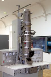 Electron microscope

Electron microscopes, which use beams of electrons instead of light, are designed for very high magnification usage. Electrons, which have a much smaller wavelength than visible light, allow a much higher resolution. The main limitation of the electron beam is that it must pass through a vacuum as air molecules would otherwise scatter the beam.
Instead of relying on refraction, lenses for electron microscopes are specially designed electromagnets which generates magnetic fields that are approximately parallel to the direction that electrons travel. The electrons are typically detected by a phosphor screen, photographic film or a CCD.
Two major variants of electron microscopes exist:
- Scanning electron microscope: looks at the surface of bulk objects by scanning the surface with a fine electron beam and measuring reflection. May also be used for spectroscopy.
- Transmission electron microscope: passes electrons completely through the sample, analogous to basic optical microscopy. This requires careful sample preparation, since electrons are scattered so strongly by most materials. It can also obtain detailed information on the sample's crystallography through selected area diffraction.
Scanning probe microscope
In scanning probe microscopy (SPM), a physical probe is used either in close contact to the sample or nearly touching it. By rastering the probe across the sample, and by measuring the interactions between the sharp tip of the probe and the sample, a micrograph is generated. The exact nature of the interactions between the probe and the sample determines exactly what kind of SPM is being used. Because this kind of microscopy relies on the interactions between the tip and the sample, it generally only measures information about the surface of the sample.
Some kinds of SPMs are:
- Atomic force microscope
- Scanning tunneling microscope
- Electric force microscope
- Magnetic force microscope (MFM)
- Near-field scanning optical microscope
Point-projection microscopes
The field emission microscope, field ion microscope, and the Atom Probe are examples of point-projection microscopes where ions are excited from a needle-shaped specimen and hit a detector. The Atom-Probe Tomograph (APT) is the most modern incarnation and allows a three-dimensional atom-by-atom (with chemical elements identified) reconstruction with sub-nanometer resolution.
Other microscopes
Acoustic microscopes use sound waves to measure variations in acoustic impedance. Similar to SONAR in principle, they are used for such jobs as detecting defects in the subsurfaces of materials including those found in integrated circuits.